Impact of Surgery on Impacted Dentures in the GIT -Case Series and Review of Literature
ADVANCED RESEARCH IN GASTROENTEROLOGY & HEPATOLOGY JUNIPER PUBLISHERS
Authored by Amarjothi JMV
Abstract
Dentures are an important cause of impaction in the Gastrointestinal tract especially in the elderly. These impacted dentures may be frequently overlooked due to their radiolucency and are frequently not amenable for endoscopic retrieval necessitating surgery for retrieval of these foreign bodies. The aim of this study is to describe the type of impaction, site, consequences and time interval to therapeutic intervention including the type of intervention and outcomes after accidental swallowing of dentures in a tertiary care referral hospital and assess the same in published medical literature throughout the world. From our experience, it is seen that dentures impacted in the cervical esophagus present earlier than those impacted in the thoracic esophagus as they are more symptomatic. Leaks at the primary closure site are more common in the cervical than thoracic esophagus which fortunately are more self-limiting and easily managed than thoracic leaks.
Keywords: Gastrointestinal tract; Dentures; Radiolucency; Endoscopic retrieval; Foreign bodies; Medical literature; Thoracic esophagus
Introduction
Impaction of dentures occurs most commonly in elderly in the oesophagus. Though most are non-impacted and amenable to endoscopic retrieval, impacted dentures usually require surgical retrieval. Delay in diagnosis of impacted dentures occurs commonly and is associated with significant morbidity and mortality.
Methodology/Results
The study was carried in a prospectively maintained database in a tertiary care hospital in southern India (2008-2017) where there is a large inflow of such impacted cases for management. Also, a comprehensive study of databases like PUBMED was also carried out to look for such cases and the inference of the study was duly noted.

Our Experience

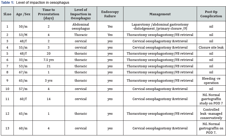
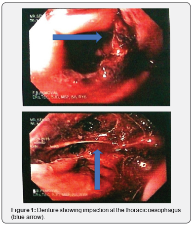
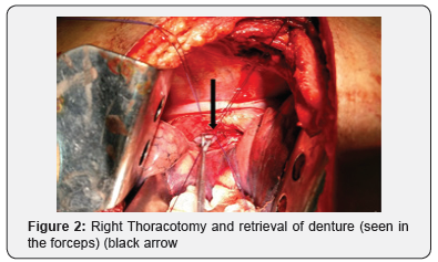
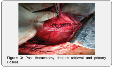
In our series, 13 patients presented (11 M:2F) (average age- 54.9 yrs) with endoscopic failed retrieval of partial, radiolucent impacted dentures without clasps at a median of 4 days (range, 1day-7.5 yrs) with 7 impacted at level of thoracic oesophagus, 5 in the cervical oesophagus and 1 in the stomach. All the impacted dentures in the cervical oesophagus were symptomatic with 71% (5/7) in the thoracic oesophagus presenting with chest pain. In view of previous failure with endoscopic retrieval, all patients with dentures impacted in the cervical oesophagus underwent cervical oesophagostomy by left cervical incision along the anterior SCM, retrieval and primary closure. All endoscopically refractory dentures impacted in the thoracic oesophagus (n=7) underwent a right thoracotomy and retrieval with intercostal drainage. The denture impacted at the OG junction was retrieved by making a gastrotomy and primary closure.40%(n=2) of patients who underwent cervical oesophagotomy and retrieval, developed leak, which was managed conservatively over a mean duration of 12 days. One of those who underwent thoracotomy (14.2%) developed leak which subsided on conservative treatment of 60 days and another patient (14.2%) developed severe bleeding after surgery necessitating a relook surgery for arresting the bleeding site. Both patients made a delayed recovery (Table 1 & Figures 1-3).


Literature Review
Incidence of impacted dentures According to current literature, the most common ingested Foreign Body (FB) in children are coins [1]. The frequency of swallowed foreign body (FB) in adults varies widely. In one study, the more commonly swallowed foreign bodies among adults are fish bones (9–45 %), bones (other than fish bones) (8–40 %), and dentures (4–18 %) [2]. Dentures are the most common FB among the elderly with a peak age incidence of 60 years [3]. The incidence of dentures as a source of impacted foreign body in the oesophagus varies widely in literature from 0.6% in a large series of over 2300 impacted oesophageal FBs to recent series which vary from 11.5% to 38.6% [4-6]. This discrepancy may be due to the sample size and reviewing of such cases from tertiary care centres only (where more difficult cases are usually referred) [7].
Dentures as foreign bodies are overlooked [8] because
a) They are irregular and allow partial passage of food, giving a false security
b) Radiolucent acrylic material is not picked up by conventional radiography.
c) Though most recent dentures lack metal wires or hooks, the few that might have, may be overshadowed by other radio opaque shadows.
The emphasis is on early removal of impacted dentures due to the following reasons: a) Chance of spontaneous passage is small, b) Oedema at the local site grips the object firmly making later manipulation increasingly difficult c) Perforation of the Oesophagus and other blood vessels may be detrimental
Dentures -types
Dentures are nowadays made of acrylic radiolucent material which is a far cry from the radiopaque metallic dentures of the 1940s [9]. which has resistance to every day wear and tear [10]. They may be of two types- complete or partial, with or without metallic clasps.
The most dangerous type of denture causing impaction is the partial radiolucent acrylic denture without clasps which due to its small size (3-4 cm) [11,7], colour and radiolucency make diagnosis by endoscopy and x rays difficult. Dentures can be classified as bridges, crowns, partial dentures and others which includes cores and fractured clasps [12]. The ingested dentures are most commonly composed of crowns followed by bridges, partial dentures, metal cores and fractured clasps [12]. It is to be noted that crowns and bridges are smaller and are more amenable to endoscopic retrieval than other types of dentures. The most common dentures associated with impaction are the upper dentures though they are the ones most amenable to retrieval due to their relatively larger size [6,7].
Pathophysiology of mastication with dentures
A removable denture is a foreign body in the oral cavity and an ill-fitting denture can have negative effects on swallowing by impairing sensation in the oral cavity and this in the elderly can be compounded by a stroke which drastically increase the risk of aspiration and dysphagia [13,8].
Level of impaction of dentures in the GIT
The level of impaction of dentures may be at either of the two Physiological constrictions (most common): The most common physiological constrictions causing impaction of swallowed dentures include
a. Hypopharynx (level of vocal cords), which is amenable to endoscopic retrieval
b. cervical oesophagus (level of upper oesophageal sphincter which is between cricopharyngeus and thoracic inlet). This is the most common site for impaction [6], and can be retrieved by both endoscopy and surgery (cervical oesophagotomy)
c. Thoracic oesophagus (level of aortic arch and left bronchus). Impacted dentures at this level are prone for life threatening complications as they are in the vicinity of the great vessels after esophageal perforation. These can be retrieved by either a thoracotomy or thoracoscopy
d. Ileocaecal region: It is the most common site for perforation [14,15], due to metallic clasps and can be managed either by laparoscopically or open surgery.
e. Sigmoid colon [16], /Rectum [17] - These [18] can be accessed either by colonoscopy or laparotomy.
Pathological strictures can also cause impaction of foreign bodies necessitating retrieval
The incidence of stricture is reported to be 66.6% for the esophageal orifice, 19% for the tracheal bifurcation, and 14.3% for the esophageal hiatus [19]. The doctrine of masterly inactivity, once the foreign body passes the physiological constrictions, the cornerstone of management of ingested foreign bodies, need not necessarily apply to dentures due to their presence of clasps, irregular shape, relatively larger size and impaction even in the distal GIT like rectum.
Clinical Features of Impacted Dentures
The need for expeditious retrieval of impacted dentures is paramount as it reported that more than 24 hrs after ingestion, the rate of complications increases from 3.2% at 24 h to as high as 23.5% after 48 hrs [20]. In a study from Nigeria, only 54.5% reported to medical centre in 48 hrs reflective of the role of late compliance as a factor in complications due to impacted dentures [6]. dentures impacted at
Cervical oesophagus
The most common clinical features of dentures impacted in the cervical oesophagus include throat pain, tenderness and pooling of saliva [6]. Other rare features include hoarseness, fever and otalgia. (< 15%). Long standing dentures in the neck can mimic malignancy and even thyroid gland [21].
Thoracic oesophagus
Most cases of impaction at the thoracic level of recent onset can present with retrosternal or back pain [7]. Dentures impacted in the thoracic oesophagus can be asymptomatic for long when they mimic a malignancy and can present suddenly with features of massive UGI bleeding due to involvement of great vessels after oesophageal perforation [22,23].
Risk Factors for Impaction of Dentures
a) Patient factors like increased risk for aspiration, epilepsy, depression, drug intake [24], late presentation to hospital, general anesthesia [25], rapid drinking pattern of liquids [7].
b) Worn out dentures, ill-fitting dentures due to bone resorption with age [6].
c) Acrylic, partial dentures
d) Strictures and spasms of the distal oesophagus [26].
Complications
Jackson [27] reported the factors that contribute to overlooking of foreign bodies:
1) Failure to consider the possibility of a foreign object when developing a differential diagnosis;
2) Absence of a history suggesting a foreign body which is common in the elderly with dentures. Factors like neurological impairment, stay alone, absence of caregivers etc. may impair an accurate history; and
3) mimic of other diseases as asthma, pneumonia, or tumor. especially most impacted thoracic dentures may mimic oesophageal malignancy and even rare complications due to impacted thoracic dentures like vocal cord paralysis, bronchial and aortic involvement may mimic complications due to oesophageal carcinoma [23].
Complications of impacted dentures can be described at the Level of oesophagus
i. Aortic erosion [22]
ii. Broncho aortic fistula [23]
iii. Horners syndrome [28]
iv. Oesophageal migration with diverticulum [29]
v. Oesophageocarotid fistula [6]
vi. RLN palsy. which is an entrapment neuropathy due to FB induced perioesophageal fibrosis [30].
vii. Tracheoesophageal Fistula (TEF) [31-33] Below the level of the oesophagus
a) Enterocolonic fistula involving small bowel and transverse colon [34].
b) Illeal impaction and perforation [35,36].
c) Rectosigmoid perforation [16-18].
Investigations -Impacted Dentures
Radiographs
Radioluscent dentures are rarely seen on lateral x-rays. Also, it is to be noted that there is a decrease in the size of the foreign body on radiological examination [7]. And hence, they do not significantly impact on subsequent management [7,37]. The classical findings include prevertebral soft tissue shadow (45%), and air entrapment around the denture (40 %) and wire clasps (27%) [6].
CT
The findings include mildly hyperdense curvature and air around the dentures may be seen [38].
Endoscopy
Rigid endoscopy: under ETGA/muscle relaxation can be done with success rate of 80% [39]. However, it is to be noted that endoscopic retrieval of dentures is associated with lacerations of the mucosa which are prone to perforation. Endoscopy, in some cases of impacted dentures, is more or less a blind procedure as vision is obscured by edematous mucosa, hidden or perforated denture edges and imperceptible hue of denture from surrounding mucosa
Manevours used to cause disimpaction and increase yield of endoscopic retrieval
a) Grasping forceps are most commonly used to retrieve dentures endoscopically [40]. In a study from Japan, Mazuno [12] described their success rate at > 90%. However, it must be noted that 13/23 (56.25%) were crowns which are smaller and amenable to easier endoscopic extraction
b) Retrieval nets can also be used to retrieve small dentures like crowns [12]. However, it must be used judiciously since their irregular surface may injure the mucosa during retrieval
c) Shear forceps to fragment and dislodge dentures and screw into the substance of FB to increase purchase before extraction [40].
d) Use of overcovering plastic incubators before extraction [40]
e) Hd YAG laser to fragment denture [41,42].
f) Oral side balloon or transparent cap to disimpact foreign body [43].
g) Cotton sliver soaked in saline to disimpact [44].
h) Long rotation of scope or sounding the foreign body with cannula in case of suspicion [7].
Complications related to perforation during endoscopic instrumentation include paraesophageal abscess, mediastinitis, pericarditis, pneumothorax, pneumomediastinum, tracheoesophageal fistula, and vascular injury [41].
Flexible fibreopric oesophagoscopy: are for use under topical anesthesia, permitting safer inspection, biopsy and photography. The standard technique using rigid oesophagoscope are not superseded, nor are they likely to be, in the foreseeable future, for foreign body removal [8].
Fluoroscopy
Upper gastrointestinal tract radiography using a nonionic water-soluble contrast medium may be used to rule out perforations (especially when aspiration is a risk) [7] (Table 2).
Treatment Options

Conservative management
There is a role for conservative management in the treatment of ingested dentures. The indications include asymptomatic patients, nonimpacted dentures, preferably small (,2.5 cm in diameter, < 6 cm in length, [45], radio dense dentures without clasps or irregular edges. The exact duration for such management is unknown which may vary from 1 -6 weeks in literature [45,46]. The advantage of such an approach is the absence of morbidity due to an additional intervention which must be balanced against the need for routine confirmatory radiography and possibility of perforation during the waiting period.
Endoscopic retrieval
The range of foreign bodies able to be removed with a flexible gastroscope has expanded with the development of newer snares, cages, and forceps. Removal of a foreign body with the flexible gastroscope is more likely to be successful and atraumatic when the foreign body does not have sharp or penetrating edges.
Glucogon
Glucagon has been used in impacted oesophageal dentures which relaxes the esophageal smooth muscle effecting the transition into the stomach. Ferrucci et al, [47] reported good outcomes using this method. However, other authors have not found glucagon to be as reliable, with success rates between 29- 50%. It is believed that use of smooth muscle relaxants/ promotility agents is contraindicated for removing all foreign bodies except food boluses [48].
Surgery
Cervical oesophagus
Cervical oesophagotomy
Since Markoe [49] demonstrated the feasibility of using the cervical approach for esophagotomy to remove foreign bodies from the esophagus, many articles have supported this as the gold standard in impacted cervical oesophageal foreign bodies in general and dentures, especially in those with failed endocopic retrieval [40,50-52]. After surgical retrieval, the oesophagostomy can be closed primarily or over a T tube in case of friable tissue [53].
Thoracic oesophagus
Posterolateral Thoracotomy over the right side is used to access the oesophagus for retrieval of impacted dentures where the oesophagus may perforate during retrieval increasing the morbidity and mortality of the procedure.
Moghissi [54] reported that ten of 39 cases with oesophagus perforation occurred during removal of the foreign body. This author reported a mortality rate of 48% in cases of thoracic oesophageal perforation. Some authors have described thoroscopic approach to access the oesophagus with reduced morbidity and mortality [56,57].
Stomach
Both open gastrotomy and laparoscopy have been tried to retrieve denture in the stomach after endoscopic failure where gastrotomy can be tried after failed laproscopic retrieval [57].
Ileocecal region/rectosigmoid
Ileocaecal region is the most frequent site of perforation especially when the swallowed object has sharp edges like clasps of a removable denture If the dentures become impacted at these sites, urgent laparotomy and Foreign body retrieval is to be done to avoid perforation.
Prevention of Impaction
A survey of dentists in the U.S. revealed that identification and retrieval of dental prostheses are complicated by the radiolucency of the materials used in the manufacture of some devices. Radiolucency in denture materials has been addressed repeatedly by dental organizations with no standard manufacturing protocol [58,59].
The mandatory incorporation of radiopaque material in dental resins is still not feasible, as they cannot match the physical properties, appearance and the ease of handling of currently used radiolucent dentures. The incorporation of heavy metal salts or glass fillers not only is unsightly, but also weakens the material, thereby increasing the risk of fracturing and swallowing a fragment [5]. Use of 12% barium fluoride [60] which maintains the mechanical and aesthetic properties and radiopaque wires have not seen widespread use [5, 61-84] (Table 3).

Conclusion
A careful clinical history and a radiologic examination is paramount in a suspected case of ingestion though it might not be very useful. Partial radiolucent dental prostheses without metal clasps, though not as common as those metal clasps to get impacted, present a diagnostic challenge. Impacted dentures the esophagus for greater than 24 hours cause oesophageal damage necessitating rapid intervention to avoid perforation. Endoscopic retrieval can be tried as first method for extraction. If the impacted foreign body cannot be removed endoscopically, surgical removal should be undertaken. Preventing the ingestion of dental prostheses involves patient education (61) regarding the potential risks of wearing broken or defective dentures.
For more articles in Advanced Research in Gastroenterology & Hepatology please click on https://juniperpublishers.com/argh/index.php
For more about Juniper Publishers please click on: https://juniperpublishers.com/video-articles.php


Comments
Post a Comment