Endoscopic Drainage of Walled off Necrosis - A "Multidisciplinary Affair"?_Juniper Publishers
ADVANCED RESEARCH IN GASTROENTEROLOGY & HEPATOLOGY JUNIPER PUBLISHERS
Authored by Zaheer Nabi
Case Report
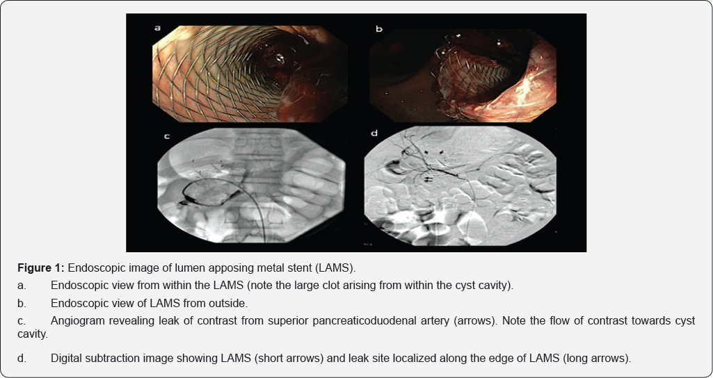
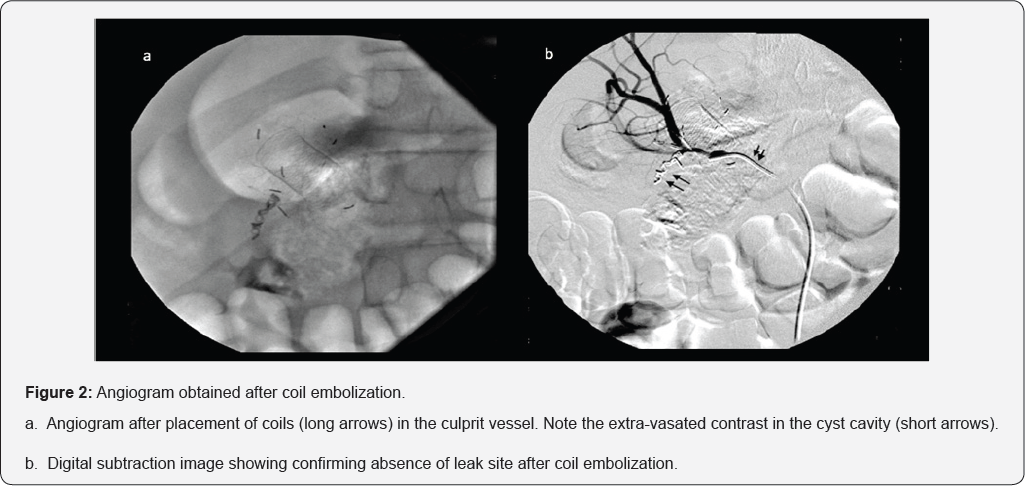
A 12-year old boy was hospitalized with history of acute necrotizing pancreatitis (7 weeks). Evaluation revealed large walled off necrosis (WON). The child had persistent symptoms in form of pain abdomen and intolerance to oral diet. Endoscopic ultrasound (EUS) guided drainage was performed using a lumen apposing metal stent (LAMS). The drainage procedure was accomplished without any immediate untoward consequences. There was significant improvement in clinical symptoms as well as the size of WON on day 3 of drainage. However, the child had a large bout of hematemesis on day 4 of drainage. Gastroscopy revealed LAMS in situ and partially occludedby a large clot arising from the cyst cavity (Figure 1a & 1b). Urgent angiography was performed via right femoral artery, which revealed leak from superior pancreaticoduodenal artery (Figure 1c &1d). The leak of contrast was localized along the lateral edge of LAMS, near farther end. Coil embolization was done following which there was no extravasation of contrast from the artery involved (Figure 2a & 2b).


Intra-procedural bleeding can be largely evaded by EUS guidance which avoids intervening vessels [1,2]. In contrast, it may not be possible to prevent delayed bleeding i.e. 3-5 weeks after endoscopic drainage. The management of WON is therefore, a multidisciplinary affair involving interventional radiologists and surgeons [3].
To Know More About Advanced Research in Gastroenterology &
Hepatology Journal
click on:
https://juniperpublishers.com/argh/index.php
To Know More About Open Access Journals Please click on: https://juniperpublishers.com/index.php


Comments
Post a Comment