Adenomatoid Odontogenic Tumor with Radicular Cyst-A Rare Presentation
Authored by Kafil Akhtar
Abstract
Adenomatoid odontogenic tumor is a benign, slow
growing lesion. It is a relatively uncommon lesion which mainly affects
females in their second decade of life, exhibiting predilection for the
anterior region of the maxilla. The lesion is usually associated with
the crown of an unerupted tooth, most commonly the maxillary canine. We
present a case of adenomatoid odontogenic tumor associated with a
dentigerous cyst affecting the left maxillary region in a 18-year-old
female, who presented with gingival swelling. Histopathologic
examination showed round to plump to spindle-shaped epithelial cells
forming aggregates with minimal connective tissue, and cuboidal or low
columnar cells forming glandular duct-like structures. The diagnosis of
adenomatoid odontogenic tumor should be considered in a case with a
corticated radiolucency in the anterior jaw, especially in teens and
young adults. The patient has shown no signs of recurrence after eleven
months of follow up period.
Keywords:Adenomatoid odontogenic tumor; Odontogenic cyst; Odontogenic tumorIntroduction
Adenomatoid odontogenic tumor (AOT) is a
slow-growing, rare oral lesion that arises from the odontogenic
epithelium. It accounts for 2.2-13% of all odontogenic tumors [1,2]. It
is a benign tumor that involves the anterior region of the maxillary
bones, with a female preponderance. Usually occurs in the second decade
of life and may be associated with odontogenic cystic lesions.
There are three variants of AOT: follicular, extra
follicular and peripheral. The follicular type is a central intra-bony
lesion associated with an unerupted tooth, which accounts for about 70%
of all cases. The extrafollicular type is also an intra-osseous lesion,
but unrelated to an unerupted tooth and represents 25% of all AOTs. The
peripheral type is a rare form that arises in the gingival tissue [2,3].
All three variants have the same histological aspect and clinical
behavior [4]. The present study reports an uncommon case of adenomatoid
odontogenic tumor, associated with a dentigerous cyst.
Case Summary
An 18 year old female presented to the surgical
clinics with a tense mass in the oral cortical bone of her maxilla for
the last 7months. Intra-oral physical examination revealed a firm,
sessile, grey-brown, 3 x 2 cm swelling in the anterior right maxillary
gingiva, with a focal, well-demarcated red-colored area in the region
adjacent to the marginal gingiva. Panorama radiographic study revealed a
well circumscribed radiolucent lesion with few radiopaque areas around
the canine tooth. An intraoral surgical intervention from the alveolar
and left central superior incisive gingiva was cARGHied out, with
removal of the tumor mass and the unerupted canine.
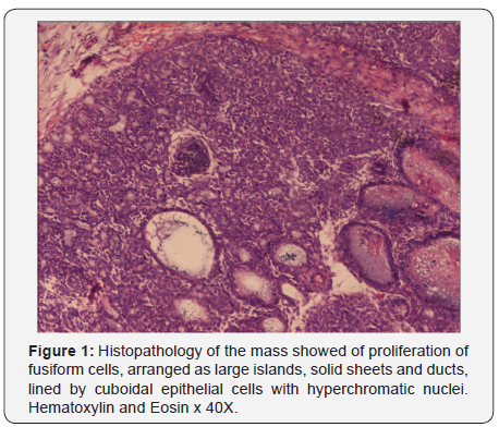
Histopathology of the mass showed of proliferation of
fusiform cells, ARGHanged as large islands, solid sheets and ducts,
lined by cuboidal epithelial cells with hyperchromatic nuclei (Figure
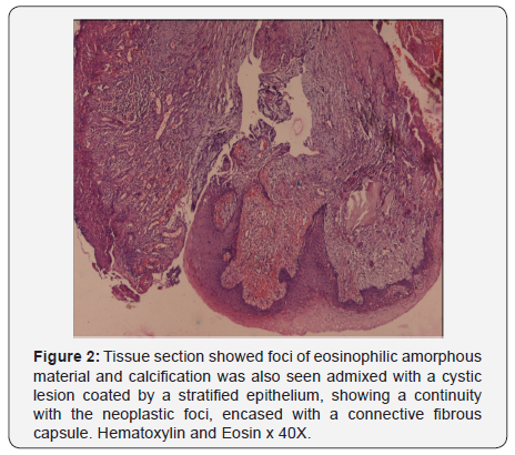
1). Foci of eosinophilic amorphous material and calcification was also
seen admixed with a cystic lesion coated by a stratified epithelium
(Figure 2), showing a continuity with the neoplastic foci, encased with a
connective fibrous capsule. A histological diagnosis of adenomatoid
odontogenic tumor, associated with a dentigerous cyst was given. After
12 months of follow up, the patient showed no signs of clinical or
radiographic evidence of tumor recurrence.


Discussion
AOT is a benign, slow growing, non-invasive odontogenic
lesion. It is rightfully called as ‘Master of Disguise’ and ‘Perfect
imitator of dentigerous cyst’ as noted in the present case [1-7].
Majority of AOTs are diagnosed in the second decade of life, with
age range of 3 to 28 years and a mean age of 13 years [3,4]. AOT
has a female preponderance with a female to male ratio of 2:1.5
AOT has been described as “ two thirds Tumor” because two
third cases occurs in the maxilla of young females and two third
cases are associated with unerupted canine tooth [4]. Our case
was seen in anterior maxilla associated with unerupted canine
tooth.
All AOT variants have a common origin as they all show an
identical histologic characteristics [6]. The pathogenesis of an
odontogenic tumor appears to involve persistence of remnants
of the dental lamina, especially after odontogenesis of the
successional and accessional laminae. These remnants give rise to epithelial rests that proliferate in response to an unknown
stimulus [7].
The radiographic findings of AOT frequently resemble
other odontogenic lesions such as dentigerous cysts, calcifying
odontogenic cysts, calcifying odontogenic tumors, maxillary
cysts, ameloblastomas and odontogenic keratocysts [8,9].
Intraoral periapical radiographs in AOT show focal radiopacities
with discrete foci of radiolucency [10,11]. The radiographic
findings in our case is typical of follicular variant of adenomatoid
odontogenic tumor, presenting as a well-defined radiolucent
unilocular lesion, associated to the crown of the unerupted tooth.
Few cases of adenomatoid odontogenic tumors associated
with dentigerous cysts are reported in the literature. Santos et
al. [5] reported a case of adenomatoid odontogenic tumor being
developed in the fibrous capsule of the dentigerous cyst. Garcia-
Pola et al. [4] described the proliferation of an adenomatoid
odontogenic cyst in the epithelial border of a dentigerous cyst.
We also noted proliferation from the cystic epithelial border and
the fibrous capsule, as described by the aforementioned authors.
Immunohistochemically, the classical AOT shows positive
staining with CK5, CK17, CK 19 and negative for CK 10, 13 and
18.11,12 Recently, Crivelini et al. [12] detected the expression of
cytokeratin 14 in AOT and concluded its origin from the reduced
dental epithelium which is also positive for cytokeratin 14
antibodies.
Conservative surgical enucleation with removal of the
unerupted dental element is the treatment modality of choice,
because of its low tendency to recur [4,5]. A more conservative
approach to treatment is marsupialization of dentigerous
cyst associated adenomatoid odontogenic tumor, and allow
the later eruption of the dental element. Recurrence of AOT is
exceptionally rare. Only three cases of recurrence in Japanese
patients have been reported [5]. Therefore, the prognosis is
excellent. No recurrence was seen in the present case.
To conclude, AOT is a relatively uncommon distinct
odontogenic neoplasm and is rightfully called as ‘Master of
Disguise’ and ‘Perfect imitator of dentigerous cyst’. It should be
a part of differential diagnosis in any oral mass lesion in a young
patient with unerupted tooth.
To Know More About Advanced Research in Gastroenterology &
Hepatology Journal
click on:
https://juniperpublishers.com/argh/index.php
https://juniperpublishers.com/argh/index.php


Comments
Post a Comment