Unusual Complication of ERCP in a Child with Bile Duct Calculi_Juniper Publishers
ADVANCED RESEARCH IN GASTROENTEROLOGY & HEPATOLOGY JUNIPER PUBLISHERS

Authored by Zaheer Nabi
Description
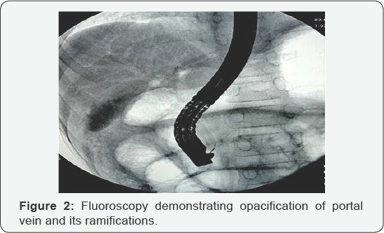
An11-year boy, presented with recurrent episodes of pain abdomen for two months. Evaluation revealed elevated serum transaminases level (ALT-118, AST-230 IU/L, upper limit of normal -40) and alkaline phosphatase level (580 IU/L). Ultrasound abdomen revealed multiple calculi in gall bladder and dilated common bile duct (CBD). Magnetic resonance cholangiopancreatogram confirmed the presence of few small calculi in CBD along with gall bladder stones. Endoscopic retrograde cholangiography was performed under deep sedation in left lateral position. Cholangiogram revealed multiple small calculi in bile duct (Figure 1). Endoscopic biliary sphincterotomy was done and balloon sweep attempted to clear CBD. However, the guide wire could not be retained in position and was inadvertently displaced out. Biliary cannulation was reattempted. Significant resistance was encountered during the passage of guide wire into CBD. On injection of small volume contrast, the CBD could not be opacified. Unexpectedly, portal vein ramifications were visualized on fluoroscopy (Figure 2). The guide wire was immediately withdrawn and biliary cannulation achieved after readjusting the position of sphincterotome guide wire complex. A double pigtail stent (7Fr, 7cm) was placed temporarily. There was no bleeding or any other untoward consequences as a result of inadvertent portal vein cannulation.

For more articles in Advanced Research in Gastroenterology & Hepatology please click on https://juniperpublishers.com/argh/index.php
For more about Juniper Publishers please click on: https://juniperpublishers.com/video-articles.php


Comments
Post a Comment