Transient Elasto graphy- The Newer Technique for Noninvasive Assessment of Liver Fibrosis_Juniper Publishers
Authored by Sudhir J Gupta
Introduction
Fibrosis is a wound healing response to injury and is
a complex dynamic process involving fibrogenesis and fibrolysis.
Chronic viral hepatitis or steatohepatitis leads to fibrogenesis through
increased synthesis of extracellular matrix components such as
collagens and glycoproteins. Assessment of fibrosis stage or the
presence of cirrhosis will often dictate treatment options as well as
provide an overall prognosis for patients with chronic liver disease of
any etiology. Historically, liver biopsy has been the primary means of
identifying fibrosis and monitoring for disease progression. However,
liver biopsy is a painful, expensive, and invasive procedure with risk
of potential complications [1].
The accurate evaluation of fibrosis using liver biopsy is also
complicated by sampling error and inter observer variation in staging of
fibrosis [2].
Given the risks of the procedure, the limited static and cross
sectional information provided in relation to overall disease
progression, as well as the error rate, the development of noninvasive
and reliable means of evaluating for the presence of fibrosis and
fibrogenesis has been an important area. Noninvasive tests of fibrosis
in viral hepatitis now include a combination of serologic markers as
well as imaging modalities [3].
As we progress into the era of safe and effective directly acting
antiviral therapy for chronic hepatitis C (CHC), various serum
biomarkers and imaging methods are now being validated for assessment of
liver fibrosis [4].
Principle of Transient Elastography
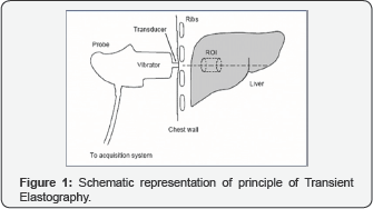
Using an ultrasound transducer probe, vibrations of
mild amplitude and low frequency (50 Hz) are transmitted through the
liver tissue (Figure 1).
This results in an elastic shear wave that propagates through the
underlying liver tissue. The probe then utilizes pulse-echo ultrasound
to follow the propagation of the shear wave and to measure its velocity.
The velocity of the wave is directly related to tissue stiffness which
correlates with fibrosis [5].
The harder the tissue, the faster the shear wave propagates. The liver
stiffness is calculated from velocity and expressed in kilopascal (kPa).
This method allows for the evaluation of numerous parameters including
velocity of vibration, velocity of wave propagation and elastic modulus.
TE allows for the identification of disease severity due to altered
mechanical properties of the fibrotic liver [6].

The feasibility and accuracy of liver stiffness
measurement (LSM) using transient elastography device are heavily
influenced by obesity and other associated factors (e.g., thoracic fold
thickness, waist circumference, and the distance between the skin and
liver capsule). Moreover, subcutaneous adipose tissue may lead to
overestimation of liver stiffness [7].
As a result, a novel probe (the 'XL' probe) is approved for use in
obese patients. This probe has a greater vibration amplitude and
measurement depth compared with the standard M probe. The XL probe
allows for accurate measurement of LSM in a significantly greater number
of obese patients than them (9-mm probe) probe [8].
In smaller adults, the standard M probe (with a 9-mm tip diameter) may
inaccurately assess liver stiffness because the nARGHow inter costal
spaces in these patients. Thus, a smaller probe, the S2 probe, has been
developed for pediatric use and, in a recent published trial by Pradhan
et al. [9]
the S2 and M probes were comparable with respect to reliability and
accuracy of stiffness measurements. However, the S2 probe may over
estimate liver stiffness in some patients, particularly those with a
larger distance between the skin and liver capsule and should be
reserved for smaller, lean patients or children.
Since fat affects the propagation of the ultrasound
wave, the transient elastography can also be used to estimate liver fat
content. This novel parameter, named Controlled Attenuation Parameter
(CAP), measures the ultrasound attenuation at the center frequency of
the M probe. In a preliminary retrospective study CAP assessment in 112
patients with chronic liver disease from various etiologies has shown
good performances for the detection and semi quantification for
steatosis [10].
Procedure of Transient Elastography
TE is a very simple and safe technique that takes
5-10 minutes and can be done in a specialty clinic or outpatient
setting. The only preliminary preparation required is that patients fast
for 2-3 hours prior to the procedure due to the potential increase in
liver stiffness from postprandial blood flow [11].
The patient is placed in a dorsal decubitus position with the right arm
in maximal abduction. The exam then begins with placement of the probe
along inter costal space to obtain a view of the right lobe of the
liver. Once an area of at least 6 cm thick and free of large vascular
structures or gallbladder has been identified, ten measurements are
obtained using the probe. The actual area measured by the probe has a
volume that is at least 100 times bigger than the average liver biopsy
sample [12].
A reliable exam should result in ten measurements
with a 70% success rate, and the inter quartile range should be less
than 30% of the value of the median [13].
An important aspect to any new technique is its cost effectiveness. TE
has been found to be a cost-effective surveillance strategy to evaluate
for the presence of fibrosis.
Limitations of Transient Elastography
TE cannot be used in individuals with ascites, and is
associated with higher failure rates or unreliable results in obese
patients using the standard M probe, as the shear wave does not
propagate through fluid, and fat also attenuates ultrasound and elastic
waves. Newer XL probes have been developed that reduce failure rates in
obese patients. Fibrosis thresholds are lower than the standard M probe,
and further validation in larger cohorts of chronic liver disease
patients is required. Children and lean patients with nARGHow inter
costal spaces also have higher failure rates, and newer pediatric S2
probes are now available to improve reliability in this regard. Liver
stiffness values for TE may be 1.3-3 times higher in the setting of
acute inflammation and/or moderate alanine amino transferase (ALT)
elevation [14].
The stiffness values usually return to baseline along with the
normalization of laboratory abnormalities. Hence, the use of TE in the
setting of trans aminitis is not recommended. Roulot et al showed that
men and patients with a body mass index >30 kg/ m2 had higher liver
stiffness scores on average. After adjusting for sex and body mass
index, liver stiffness values were also higher in subjects with
metabolic syndrome [15].
Other limitations for accurate stiffness reading include sinusoidal
congestion, extra hepatic cholestasis, TE is somewhat operator
dependent. Therefore, there may be some variability in results depending
on the operator. Utility of transient elastography in Hepatitis C,
Hepatitis B, Nonalcoholic fatty liver disease, Cholestatic Liver Disease
is already increasing.
Conclusion
Transient Elastography provides a non-invasive and
reproducible tool which can be easily utilized for patient care as an
adjunct to clinical evaluation for the staging of liver fibrosis. The
key clinical parameter in patient management is the diagnosis or
exclusion of advanced fibrosis and cirrhosis. The transient elastography
is an adjunct to clinical, radiological, and biochemical evaluation and
not a replacement to the liver biopsy When elastography does not
correlate well with clinical findings, use of serological tests such as
APRI, Hepascore, and FibroSure/ FibroTest and liver biopsy should be
considered. Utility of transient elastography in Hepatitis C, Hepatitis
B, Nonalcoholic fatty liver disease, Cholestatic Liver Disease is
already increasing.
To Know More About Advanced Research in Gastroenterology &
Hepatology Journal
click on:
https://juniperpublishers.com/argh/index.php
https://juniperpublishers.com/argh/index.php


Comments
Post a Comment